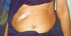
| SINGLE HOLE SURGERY FOR GALL BLADDER STONE |
| Gall bladder is surrounded by several structures. Injury to these structures can be morbid or life threatening. Membranes cover several of these structures (adhesions that form due to disease also obscure them). Thus these structures are not visible to the naked eye or through 'scopes' until & unless dissection is carried out. Unfortunately these structures are not placed similarly in all individuals & a number of variations occur, leading to the chance of injury during dissection. The only way to know about the placement of these structures before dissection is by performing ultrasound during surgery & keeping the ultrasound probe on the area to be dissected. |

| AIM OF THIS SURGERY |
examination of body gives information about structures and diseases lying deep within. When an ultrasound probe is put through the abdominal wound
inside and placed in contact with the body parts (intraoperative ultrasonography), information can be obtained about the structure / disease /
inside & contents / relations to surrounding structures of that part. Thus putting in of an ultrasound probe inside the body during surgery
gives informations which otherwise can be obtained only by touch. This information cannot be obtained by "sight" no matter how powerful
'scopes' are used. Truly speaking "intraoperative ultrasonography" provides information much more than what touch can give when used in context of
gall bladder surgery. In traditional or key-hole surgeries dye is injected inside the bile duct to detect stones but this involves risk of reactions, is
technically difficult, time consuming and many times not done routinely. In this intraoperative ultrasonography the bile duct is scanned by an ultrasound
probe thus there is no need to use the dye, once learned it is technically simple & less time consuming. Most important is the role of intraoperative
ultrasonography in identifying and saving wrong structures from being cut. Before dissection the ultrasound probe is placed on the part to identify it if
there is any doubt. In addition to intraoperative ultrasonography this SINGLE HOLE SURGERY uses Laparoscope to magnify structures as &
when needed. This use is done without filling any GAS in abdomen - routine 4 hole laparoscopic surgery for gall bladder requires filling of gas in
abdomen which has its own dangers. This use of laparoscope in single hole surgery aids "sight". During this surgery the surgeon is directly looking at
the organ he is operating upon thus the surgery is under natural 3 dimensional view. But In 4 hole laparoscopic surgery the surgeon is looking at the
monitor while operating on the patient, this is a 2 dimensional & indirect view - thus it makes surgery difficult unnecessarily as it requires guessing of
third dimension as well as unnatural hand eye coordination has to be practiced, a sort of a circus. With an aim to provide both "improved Sight"
and " advantages of Touch" in a minimally invasive surgical technique for gall bladder surgery, this technique of LAPAROSCPIC
ASSISTED INTRAOPERATIVE ULTRASOUND GUIDED SINGLE HOLE CHOLECYSTECTOMY (LAIOUSC) was developed.
'SIGHT' and 'TOUCH' are the two most essential of the senses that play their role in surgery. In "Minimally
Invasive Surgery" while Sight got priority inform of "Scopes" and "Improved illumination techniques", Touch was
left neglected. It can be appreciated that in any type of 'key- hole operations' surgeon's hand cannot enter the
abdomen, whether it is a single-hole or four-hole surgery. Hence "touch"sensation & its advantages cannot be
utilised by the surgeon. In gall bladder surgery (cholecystectomy) "Touch" provides safety to the bile duct system
against injury & prevents leaving behind of stones & parts of gall bladder to an appreciable extent. Ultrasound
